
Larry Gordon is a 1.5-meter-long eel who needs a CT scan
New York Times

- +
- -
Larry Gordon had a lump.
Larry, 30, a leopard moray eel, was a staff and visitor favorite at the Point Defiance Zoo & Aquarium in Tacoma, Washington. The 1.5-meter-long fish had a spirited personality — he had been rumored to hold grudges against aquarists who had fallen into his disfavor — and a punk-rock look, with a spotted dorsal fin running down his spine like a well-gelled mohawk. Like other moray eels, he had two sets of jaws and an abundance of teeth, which he used to rip into the fish fillets doled out for dinner.اضافة اعلان
But this spring, he stopped eating, and veterinarians soon found a fleshy pink growth on the roof of his mouth.
They found a broken tooth, too, which they initially thought might explain Larry’s oral misfortune. When they extracted the offending fragment, the lump vanished, and the eel started eating again.
“He was feeling better,” said Dr Kadie Anderson, an associate veterinarian at the zoo. “And I was like, ‘Great, we’ve got this behind us.’”
Then, this fall, the mysterious mass of cells grew back, and Anderson began to worry that Larry might have a larger tumor lurking somewhere in his skull. Thirty is old in eel years, and an invasive cancer might necessitate euthanasia, she said.
To determine whether the mass extended into Larry’s nasal passages or bone, veterinarians would need to give him a CT scan. The scans are not invasive, but imaging an eel poses a slippery challenge, requiring zoo employees to ferry Larry to a veterinary hospital across town, safely hoist him out of the water, and somehow get him into a fancy piece of medical equipment.
“And saltwater does not play well with electronics,” Anderson said.
They would need to be careful, they would need to be fast, and they would need to remember the baking soda.
Here is how they got the job done:
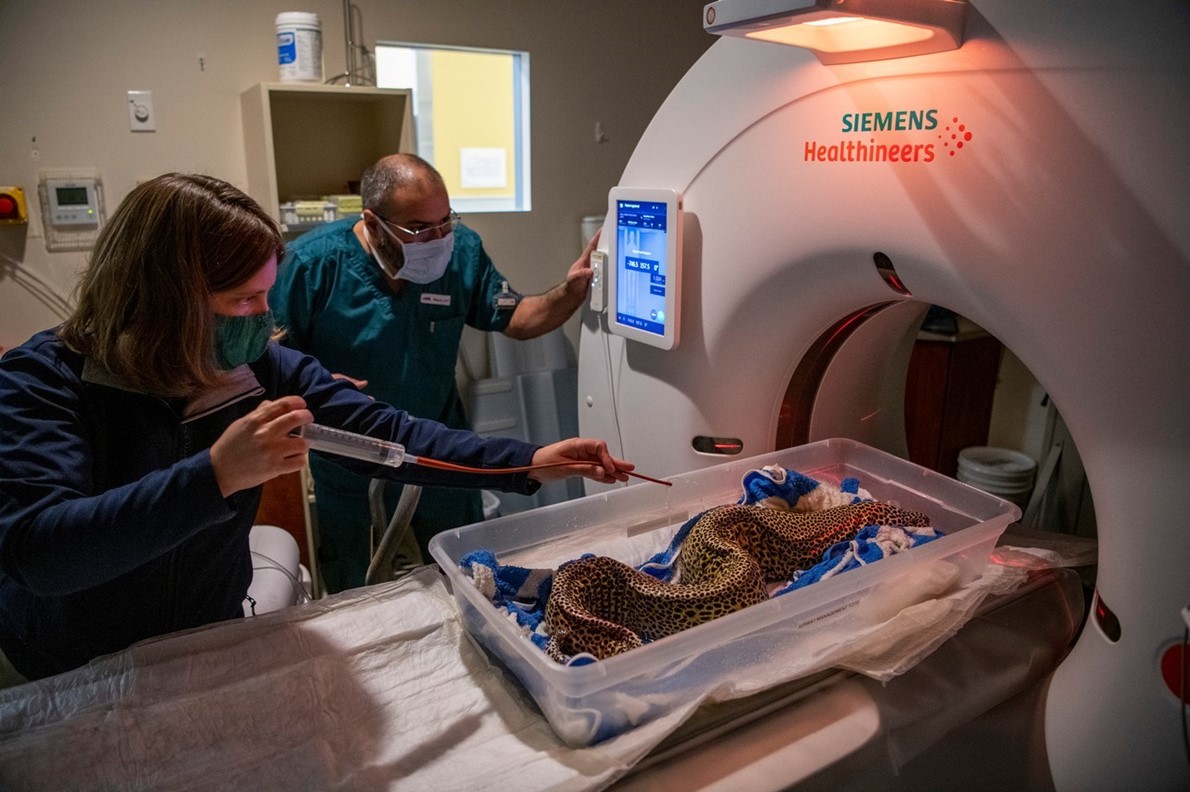
Dr Kadie Anderson, an associate veterinarian at the Point Defiance Zoo & Aquarium, streaming medicated water onto Larry Gordon, a 30-year-old leopard moray eel, before his CT scan as radiology tech Miguel Contreras-Avelar looks on
The logistics of fish transportation come down to good water management. In Larry’s permanent habitat, the water temperature and oxygen levels were carefully calibrated.
“But when you’re transporting him, you don’t have all that life support,” Anderson said.
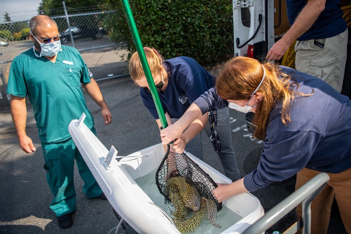
So zoo staff loaded Larry, a warm-water fish, into a big plastic tub in the back of a heated van, bubbling oxygen into the water during the 20-minute drive to Summit Veterinary Referral Center. Upon arrival, they transferred him to an insulated, water-filled cooler and wheeled him into the hospital.
Despite his age, Larry remained an active eel. To ensure that he did not harm himself, or the scanner, the veterinary team needed to immobilize him.
“When humans go through CT, you can tell a person to hold still,” Anderson said. “I can’t make an eel hold still.”
So they added a powdered anesthetic, known as MS-222, to the water in the cooler. The drug, which fish can absorb through their gills or skin, works quickly but also makes the water acidic. To keep the pH level steady, and Larry safe, the veterinary team also added a dash of baking soda.
Within a few minutes, Larry was under. Zoo employees lifted all 16 pounds of him from the cooler, cradling him in a wet towel to protect his mucus-covered skin and flexible spine. (Like dachshunds and other improbably long animals, eels are prone to back problems, Anderson said.) Then they gently laid him in a shallow plastic bin, the kind familiar to many storage-strapped college students.
“You know those Tupperwares that you can slide under your bed?” Anderson said.
As the staff members placed Larry in the container, they tried to gently unfurl his serpentine body.
“When you do a CT, you want to not have things all folded up and overlapping,” Anderson said.
Even then, though, the eel was too long to straighten completely. Anderson used a rubber catheter to stream more medicated water onto Larry’s skin and gills.
“We can do surgery on fish out of the water as long as you have their gills getting perfused with water,” she said. “It’s like putting a breathing tube into a person.”
And then they slid the plastic bin into the doughnut-shape scanner, which bombarded the eel with X-rays from multiple angles, generating detailed images of Larry’s internal anatomy.
The images bore good news: Larry’s lump did not look like cancer and had not infiltrated the bone. Veterinarians were able to review the images immediately, but the radiologist needed some time to assemble her final report, Anderson said. “She never imaged an eel before.”
To rouse Larry, the team deposited him in a cooler filled with warm, drug-free water. Within a few minutes, he had righted himself — anesthetized fish often list to the side — and was moving around again. Then, he hitched a ride home in the heated van.
The CT scans convinced Anderson that she could safely remove the rogue mass from Larry Gordon’s mouth, a procedure she recently completed. The eel is now healing, she said, and the image of his sinuous skeleton has become a “beautiful” keepsake, perhaps even one that the zoo could put on display.
“I think a 3D reconstruction of the eel would be really neat to share with visitors,” Anderson said.
The image could be Larry’s legacy, teaching visitors about the remarkable anatomy of eels even after the geriatric fish, and his mysterious mass, are gone.
Read more Odd and Bizarre
Jordan News
Larry, 30, a leopard moray eel, was a staff and visitor favorite at the Point Defiance Zoo & Aquarium in Tacoma, Washington. The 1.5-meter-long fish had a spirited personality — he had been rumored to hold grudges against aquarists who had fallen into his disfavor — and a punk-rock look, with a spotted dorsal fin running down his spine like a well-gelled mohawk. Like other moray eels, he had two sets of jaws and an abundance of teeth, which he used to rip into the fish fillets doled out for dinner.
But this spring, he stopped eating, and veterinarians soon found a fleshy pink growth on the roof of his mouth.
They found a broken tooth, too, which they initially thought might explain Larry’s oral misfortune. When they extracted the offending fragment, the lump vanished, and the eel started eating again.
“He was feeling better,” said Dr Kadie Anderson, an associate veterinarian at the zoo. “And I was like, ‘Great, we’ve got this behind us.’”
Then, this fall, the mysterious mass of cells grew back, and Anderson began to worry that Larry might have a larger tumor lurking somewhere in his skull. Thirty is old in eel years, and an invasive cancer might necessitate euthanasia, she said.
To determine whether the mass extended into Larry’s nasal passages or bone, veterinarians would need to give him a CT scan. The scans are not invasive, but imaging an eel poses a slippery challenge, requiring zoo employees to ferry Larry to a veterinary hospital across town, safely hoist him out of the water, and somehow get him into a fancy piece of medical equipment.
“And saltwater does not play well with electronics,” Anderson said.
They would need to be careful, they would need to be fast, and they would need to remember the baking soda.
Here is how they got the job done:
Dr Kadie Anderson, an associate veterinarian at the Point Defiance Zoo & Aquarium, streaming medicated water onto Larry Gordon, a 30-year-old leopard moray eel, before his CT scan as radiology tech Miguel Contreras-Avelar looks on
The logistics of fish transportation come down to good water management. In Larry’s permanent habitat, the water temperature and oxygen levels were carefully calibrated.
“But when you’re transporting him, you don’t have all that life support,” Anderson said.
So zoo staff loaded Larry, a warm-water fish, into a big plastic tub in the back of a heated van, bubbling oxygen into the water during the 20-minute drive to Summit Veterinary Referral Center. Upon arrival, they transferred him to an insulated, water-filled cooler and wheeled him into the hospital.
Despite his age, Larry remained an active eel. To ensure that he did not harm himself, or the scanner, the veterinary team needed to immobilize him.
“When humans go through CT, you can tell a person to hold still,” Anderson said. “I can’t make an eel hold still.”
So they added a powdered anesthetic, known as MS-222, to the water in the cooler. The drug, which fish can absorb through their gills or skin, works quickly but also makes the water acidic. To keep the pH level steady, and Larry safe, the veterinary team also added a dash of baking soda.
Within a few minutes, Larry was under. Zoo employees lifted all 16 pounds of him from the cooler, cradling him in a wet towel to protect his mucus-covered skin and flexible spine. (Like dachshunds and other improbably long animals, eels are prone to back problems, Anderson said.) Then they gently laid him in a shallow plastic bin, the kind familiar to many storage-strapped college students.
“You know those Tupperwares that you can slide under your bed?” Anderson said.
As the staff members placed Larry in the container, they tried to gently unfurl his serpentine body.
“When you do a CT, you want to not have things all folded up and overlapping,” Anderson said.
Even then, though, the eel was too long to straighten completely. Anderson used a rubber catheter to stream more medicated water onto Larry’s skin and gills.
“We can do surgery on fish out of the water as long as you have their gills getting perfused with water,” she said. “It’s like putting a breathing tube into a person.”
And then they slid the plastic bin into the doughnut-shape scanner, which bombarded the eel with X-rays from multiple angles, generating detailed images of Larry’s internal anatomy.
The images bore good news: Larry’s lump did not look like cancer and had not infiltrated the bone. Veterinarians were able to review the images immediately, but the radiologist needed some time to assemble her final report, Anderson said. “She never imaged an eel before.”
To rouse Larry, the team deposited him in a cooler filled with warm, drug-free water. Within a few minutes, he had righted himself — anesthetized fish often list to the side — and was moving around again. Then, he hitched a ride home in the heated van.
The CT scans convinced Anderson that she could safely remove the rogue mass from Larry Gordon’s mouth, a procedure she recently completed. The eel is now healing, she said, and the image of his sinuous skeleton has become a “beautiful” keepsake, perhaps even one that the zoo could put on display.
“I think a 3D reconstruction of the eel would be really neat to share with visitors,” Anderson said.
The image could be Larry’s legacy, teaching visitors about the remarkable anatomy of eels even after the geriatric fish, and his mysterious mass, are gone.
Read more Odd and Bizarre
Jordan News